Tension pneumothorax: manifestations and treatment options
Content
- Types of pneumothorax
- What causes tension pneumothorax?
- What are the symptoms of tension pneumothorax?
- Detection and treatment options
- Implementation of pleural needle decompression
- Decompression needle
- Possible complications
- Recommendations for further medical care

A tension pneumothorax is a life-threatening condition. It develops due to the constant flow of air into the pleural cavity, which leads to lung compression and prevents their proper expansion.
It also affects the heart, reducing cardiac output, as well as blood vessels and other structures in the chest. If not treated promptly, the pathology progresses rapidly, which can cause cardiovascular collapse and, ultimately, cardiac arrest.
A feature of tension pneumothorax is damage to the pleura, which acts as a one-way valve. Because of this, air can enter the pleural space during inhalation but cannot escape during exhalation. With each breath, more air is trapped in the chest, leaving less room for the lungs to expand.
Types of pneumothorax
A pneumothorax is a condition in which air accumulates in the pleural cavity due to damage to the lungs or chest. The presence of air in the pleural cavity compresses the lung and impairs gas exchange.
Classification of pneumothorax:
1. Depending on the reason:
- Spontaneous: It occurs due to rupture of emphysematous bullae or subpleural alveoli. It can be primary (in healthy people without previous lung disease) or secondary (in diseases such as COPD, cystic fibrosis, histiocytosis from Langerhans cells, lymphangioleiomyomatosis).
- Post-traumatic: It occurs as a result of chest trauma, both with and without a violation of the integrity of the membranes (sharp object injury, falling from a height, compression, road traffic accident).
- Iatrogenic: It occurs due to medical procedures such as thoracentesis, lung biopsy (percutaneous or transbronchial), catheterisation of large veins (subclavian, rarely internal jugular), mechanical ventilation, thoracic surgery.
2. Depending on the mechanism of occurrence:
- Closed: A certain amount of air is trapped in the pleural cavity, which can resolve on its own within a few days (e.g. iatrogenic pneumothorax after a pleural puncture).
- Open: Air freely enters the pleural cavity through an opening in the chest or bronchus and freely exits through the same opening. This can cause "pendulous movements of the mediastinum" that can cause reflex cardiac arrest.
- Tight (valve): A valve forms in the opening through which air enters the pleural cavity. When you inhale, air enters the pleural cavity but cannot escape when you exhale.
Intrapleural pressure exceeds atmospheric pressure and is constantly increasing, which leads to lung compression, mediastinal displacement, compression of the other lung, large venous vessels, decreased venous outflow and cardiac output.
This causes severe hypotension and hypoxaemia, which can lead to sudden circulatory failure. A tension pneumothorax is a life-threatening condition that requires immediate intervention.
3. Depending on the size:
- Small: The distance between the chest wall and the visceral pleura (lung margin) at the level of the lung gate on a posterior-anterior chest radiograph is less than 2 cm.
- Large: Distance of 2 cm or more.
What causes tension pneumothorax?
A tension pneumothorax occurs due to specific mechanisms and causes that lead to the formation of a one-way valve in the pleural cavity. This valve allows air to enter during inhalation, but does not release it during exhalation, which causes a gradual increase in intrapleural pressure. The main causes of tension pneumothorax include:
1. Injuries to the chest:
- Penetrating injuries: Sharp object injuries (stab wounds or gunshot wounds) can damage the pleura, forming a valve mechanism.
- Blunt trauma: Chest compression or blows can cause rib fractures that can damage the pleura and lungs, creating conditions for the development of a tension pneumothorax.
2. Medical procedures:
- Mechanical ventilation: In patients who are connected to a ventilator, high ventilation pressure can cause the alveoli to rupture and air to enter the pleural cavity.
- Thoracentesis: Inserting a needle into the pleural cavity to remove fluid or air can accidentally damage the lung or pleura.
- Lung biopsy: Both percutaneous and transbronchial biopsies can cause pleural damage and pneumothorax.
3. Pulmonary diseases:
- Chronic obstructive pulmonary disease (COPD): Patients with COPD may develop emphysematous bullae that can rupture and cause pneumothorax.
- Cystic fibrosis: This disease is often accompanied by the formation of cysts and bullae in the lungs, which can rupture.
- Other rare diseases: Histiocytosis from Langerhans cells, lymphangioleiomyomatosis and other pulmonary pathologies may predispose to tension pneumothorax.
4. Spontaneous reasons:
- Primary spontaneous pneumothorax: Occurs in healthy people without apparent cause or previous lung disease, often due to rupture of small subpleural bullae.
- Secondary spontaneous pneumothorax: Associated with pre-existing lung disease that causes bullae or cysts to form and rupture.
- Intense physical activity or sudden changes in pressure: Certain activities, such as strenuous physical labour, scuba diving or flying at high altitudes, can contribute to the development of pneumothorax due to sudden fluctuations in lung pressure.
Thus, the onset of tension pneumothorax is influenced by both external factors, such as trauma and medical interventions, and internal pathological conditions of the lungs. Timely detection and treatment are critical to prevent serious complications and save the patient's life.
What are the symptoms of tension pneumothorax?

A patient with a pneumothorax experiences a sudden onset of shortness of breath accompanied by stabbing chest pain. Chest mobility is limited on the affected side.
Respiratory sounds on auscultation (listening to the chest with a stethoscope) are quieter than normal, and a drum-like sound is heard on percussion (tapping). In case of tension pneumothorax, there is increasing dyspnoea and displacement of the mediastinum, which can be detected by determining the position of the trachea above the jugular notch of the sternum.
The diagnosis is confirmed by chest X-ray, which is performed with full exhalation. Small pneumothoraxes sometimes go undiagnosed, but this is usually not clinically relevant.
In critical situations, there may not be time for an examination, and the doctor must make a diagnosis based on the symptoms. In case of a tension pneumothorax, lack of timely treatment can lead to death.
The patient's life can be saved by a pleural puncture - insertion of a tube or needle into the pleural cavity to remove excess air. A tension pneumothorax is a life-threatening emergency.
The pressure in the pleural cavity should be reduced by inserting an intercostal cannula or a large hollow needle into the pleural cavity.
Detection and treatment options
Detecting and treating pathological conditions is critical to preserving the health and life of patients. Modern medicine offers a variety of diagnostic and therapeutic methods that allow for the effective detection and treatment of a wide range of diseases.
One of the most important aspects of treatment is the timely and accurate detection of the problem, which allows you to choose the most appropriate and effective method of therapy.
Various methods are used to diagnose diseases, including clinical examination, laboratory tests, instrumental studies and imaging technologies.
Clinical examination includes examination of the patient, history taking and physical examination. Laboratory tests (blood, urine, and other biological fluids) can detect abnormalities at the molecular and cellular levels.
Instrumental examinations include electrocardiography (ECG), electroencephalography (EEG) and other methods to assess the functional state of organs and systems.
Imaging techniques such as ultrasound (US), computed tomography (CT) and magnetic resonance imaging (MRI) provide detailed information about the structure of organs and tissues.
Depending on the diagnosis and severity of the disease, different treatment methods are used:
- Conservative treatment includes drug therapy, physiotherapy, diet therapy and other methods that do not require surgery.
- Drug therapy is based on the use of medications that can be aimed at relieving symptoms, killing infections, reducing inflammation or correcting metabolic processes.
- Invasive methods include minimally invasive procedures such as catheterisation, endoscopy and other technologies that allow for diagnosis or treatment with minimal tissue damage.
- Surgical treatment involves surgical intervention to remove pathological formations, reconstruct organs or eliminate the causes of the disease.
One of the most modern and effective methods of treating certain pathological conditions is needle decompression. This method is used to relieve pressure in tissues or body cavities, which may be necessary for various medical conditions such as pneumothorax, haematomas, or intervertebral hernias.
Needle decompression can quickly reduce pain and restore organ function, making it an important tool in the arsenal of modern medicine.
Implementation of pleural needle decompression
In case of tension pneumothorax, it is necessary to decompress the chest using a needle catheter. The procedure should be performed in the second intercostal space along the midclavicular line.
Remember that the first rib is located under the clavicle. For decompression, use a catheter with a large bore and a length of at least 8 centimetres.
Treat the area with alcohol wipes and insert the needle, focusing on the third rib. To do this, press on the third rib and then move the needle slightly higher, into the second intercostal space, just above the third rib, where it passes into the pleural cavity.
The catheter is inserted over the third rib to avoid damaging a vein, nerve or artery. Insert the needle with the catheter, reach the pleural cavity, wait 10 seconds and remove the needle, leaving the catheter in the patient's body.
After needle decompression, immediately perform lung auscultation and percussion to ensure that the treatment is having a positive effect. Make sure the patient feels better.
If for some reason it is not possible to insert the catheter into the second intercostal space, it should be inserted into the fifth intercostal space. Depending on the age and size of the patient, the most likely place of insertion will be along the anterior axillary line.
Both areas are equally suitable for decompression, so they are not considered as a primary and secondary option. The main thing is to focus on the rib and insert the catheter over it.
Remember that catheters in the anterior axillary line tend to bend due to the position of the patient's arm on the stretcher. Therefore, constantly monitor the patient's condition.
If the air whistling stops, turn the catheter a quarter turn and reassess the patient. This may unbend the catheter if it has become compressed at some point.
If there is still no whistling after turning, check whether the patient is breathing more easily. If breathing is still difficult, perform another decompression. The decompression site remains the same, but move 1 cm laterally from the previous site.
Leave the catheter in place to allow air to escape and prevent recurrent tension pneumothorax. In any case, be prepared for a second decompression, as catheters can easily become pinched and clogged.
Adjust the supply of supplemental oxygen. The method and rate of oxygen supply depends on many factors.
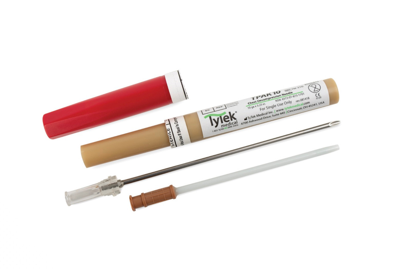
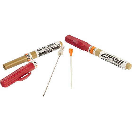
Decompression needle
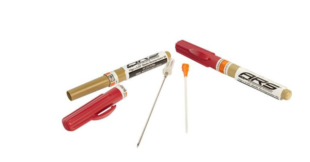
A decompression needle is an important tool in medical practice for relieving pressure in body cavities in critical conditions. This instrument is used to quickly and effectively relieve pressure in the pleural cavity (needle decompression), haematoma, or other cavities where fluids or air may accumulate, threatening the patient's life.
A decompression needle is a long and thin needle that has a wide internal channel for rapid pressure release. It can be of different lengths and diameters depending on the application and the need for medical intervention.
The main task of the decompression needle is to create a pathway for air or fluid to be removed from the body cavity, which helps to restore normal pressure and organ function.
The purpose of a decompression needle is to quickly relieve pressure in body cavities to prevent or treat critical conditions. The main applications of a decompression needle include:
- Needle decompression of the pleural cavity: Used to relieve a tension pneumothorax, where air builds up in the cavity between the lungs and the pleural membrane, which can lead to pressure on the lungs and breathing difficulties.
- Decompression of haematomas: It is used to remove blood from a subcutaneous or intramuscular haematoma, which helps to avoid further complications and facilitate the healing process.
- Other body cavities: Decompression needles can also be used to remove fluid or air from other body cavities, such as the pericardial or peritoneal cavities.
The technique of using a decompression needle involves some key steps:
- Preparation of the area: The area where the needle is to be inserted is treated with an antiseptic to prevent infection.
- Needle insertion: The needle is inserted into the body cavity using a specially developed technique, following anatomical landmarks.
- Expelling fluid or air: After the needle is inserted, the fluid or air that has accumulated in the body cavity is drained through the needle, relieving pressure and providing relief to the patient.
- Monitoring: After the procedure, it is important to provide the patient with medical monitoring to detect any complications and restore a stable condition.
The decompression needle is an important tool in rescue medicine and helps save patients' lives in critical situations. The correct use of this tool requires the knowledge of professional healthcare professionals and a high level of medical expertise.

Possible complications
Despite all the safety precautions and professionalism, the use of a decompression needle can be fraught with difficulties and possible complications. Understanding these aspects and knowing how to avoid them is critical to ensuring patient safety and the effectiveness of the medical intervention.
Difficulties with the use of a decompression needle:
- Anatomical features: Needle insertion may be difficult in the presence of abnormal anatomical features or tissue changes.
- Thick skin or muscle: In thick layers of skin or muscle, needle insertion may be difficult or impossible.
- Difficulties with cavity localisation: Some cavities, such as the pericardial cavity, can be difficult to access, making needle insertion more challenging.
- Difficulty controlling the catheter: During needle insertion, it may be difficult to control the movement of the catheter in the cavity.
Ways to avoid difficulties and complications:
- Detailed examination: Before the procedure, the anatomical features and potential difficulties of needle insertion should be carefully assessed.
- Use of additional visualisation tools: The use of ultrasound or X-ray navigation guidance can facilitate accurate needle placement into the cavity.
- Skill and experience of medical staff: It is important to have skilled and experienced medical staff who can effectively handle complexities and avoid complications.
- Sustainable training and skills development: Healthcare staff should continuously improve their skills, participate in trainings and courses to effectively cope with challenges and avoid possible complications.
- Planning and communication: Before the procedure, it is important to establish a clear plan of action and ensure effective communication between members of the healthcare team.
Avoiding difficulties and complications during the use of a decompression needle requires care, experience and cooperation of the entire medical team. Knowledge of the possible risks and methods of avoiding them are important to ensure the safety and successful outcome of the medical intervention.
Recommendations for further medical care

After a pneumothorax, especially after surgery, it takes time to fully recover and adapt. Patients may experience chest pain or discomfort for a period of time. The doctor may prescribe sedatives or recommend methods to reduce pain. Gradually, with recovery, unpleasant symptoms disappear. To control and monitor the condition of patients, the doctor will schedule regular visits.
Pneumothorax itself can be a source of emotional stress. Understanding this, patients may feel anxious, fearful or depressed. It is important to discuss your emotions with your doctor. To prevent a recurrence, the specialist will provide specific advice, which may include adherence to restrictions, regular check-ups, or lifestyle changes.
Getting accurate information about pneumothorax and discussing any questions or concerns with your doctor is important. This will help you better understand your condition and make the right decisions for your health.
Each case of pneumothorax is unique, so each patient's experience may vary. Your doctor is the best source of information and guidance about expectations after a pneumothorax and next steps.
The recovery period can vary depending on various factors such as the type and severity of the pneumothorax, the treatments used and the patient's general health. General recommendations may include:
- refraining from excessive physical activity in the initial stages;
- avoiding exertion that can increase pressure in the chest cavity;
- ensuring sufficient hydration of the body;
- giving up bad habits, such as smoking;
- healthy eating;
- maintaining a positive psycho-emotional state (if necessary, seeking psychological support);
- regular medical examinations;
- good rest and normal sleep;
- compliance with medical recommendations.
A tension pneumothorax is a medical emergency that requires immediate intervention and a comprehensive approach to treatment. Timely diagnosis, prompt and effective treatment, and preventive measures are the main components of successful management of this condition.