Haemorrhagic shock: emergency care
Content
- What is haemorrhagic shock?
- Signs and criteria for diagnosing the disease
- Conditions for the provision of medical care
- Diagnostics
- Treatment programme
- Indications for haemotransfusions
- The result of treatment
- Possible complications and side effects
- Recommendations for further treatment
- Frequently asked questions and answers
- What is haemorrhagic shock?
- What can be the causes of haemorrhagic shock?
- What are the symptoms of haemorrhagic shock?
Haemorrhagic shock is a type of hypovolaemic shock and is a critical condition that requires immediate treatment in the intensive care unit. The haemorrhagic syndrome is not an independent disease, but a non-specific pathological condition that occurs against the background of other health disorders.
It is of particular importance for medical professionals of all specialities, as it is characterised by rapid progression, severe course and unfavourable prognosis in the absence of immediate treatment.
What is haemorrhagic shock?
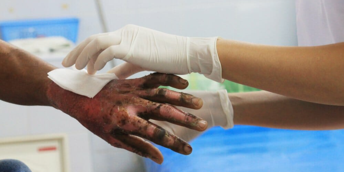
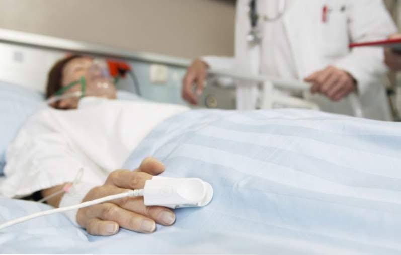
Haemorrhagic shock is a serious medical condition that occurs as a result of significant blood loss. Due to the large loss of blood, the body cannot sufficiently supply the tissues with oxygen, which can lead to organ dysfunction and, ultimately, death.
This type of shock can occur as a result of trauma, bleeding after surgery, internal bleeding due to ruptured blood vessels or organ damage, as well as massive blood loss during various medical conditions such as abdominal aortic dissection, acute pancreatitis or acute anaemia.
Symptoms of haemorrhagic shock include pale skin, cold and clammy sweat, rapid pulsing, weakness, dizziness, confusion or fatigue, and you may feel threatened by drowsiness or agitation.
Treatment of haemorrhagic shock includes restoration of blood circulation and blood loss, which may require a transfusion of blood or other blood substitutes, as well as surgery to stop the source of bleeding.
Signs and criteria for diagnosing the disease
The main factor underlying severe haemorrhagic shock is a large blood loss, exceeding 25-30% of the circulating blood volume (1.25-1.5 litres). Patients may experience thirst, impaired consciousness, pallor of the skin and mucous membranes, sunken veins, and tachycardia (heart rate of more than 100 per minute).
In elderly patients, the heart rate may remain unchanged. The pulse may be weakly filled, in severe cases (with a loss of more than 30% of the circulating blood volume) the pulse becomes threadlike or not palpable at all in the peripheral arteries.
A decrease in blood pressure is one of the key symptoms; first, there may be a decrease in pulse pressure, followed by systolic and diastolic blood pressure. Systolic blood pressure, which is lower than diastolic blood pressure, is critical for organ perfusion; normally, this figure is 75-85 mmHg. With minor blood loss, central venous pressure may remain normal, but with deepening shock, it decreases below 60 mmHg.
Hypovolaemia can be assessed by reducing the pulmonary artery wedge pressure (PAP) to less than 7 mmHg. The cardiac index may be less than 2.5 l/min/m², and peripheral vascular resistance increases. Diuresis decreases.
Laboratory parameters may also indicate haemorrhagic shock. These include anaemia, which is characterised by a haemoglobin concentration of less than 100 g/l, a red blood cell count of less than 3*10^9/l, and a haematocrit decrease of less than 0.30 l/l.
Metabolic acidosis with a pH of less than 7.3 and a base deficiency of -3 mmol/l may occur. Venous blood oxygen saturation may decrease, and blood lactate levels may increase, indicating impaired oxygen transport to tissues and its consumption.
Conditions for the provision of medical care
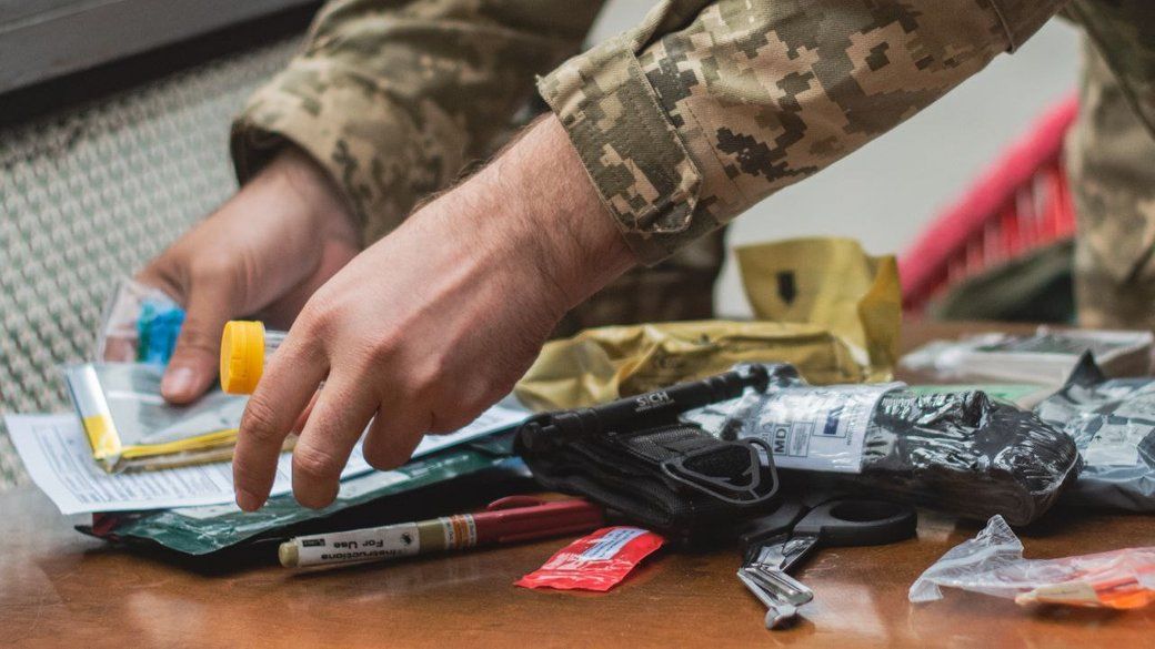
At the pre-hospital stage, it is necessary to provide urgent medical care, and in the hospital - conditions for treatment in the operating room or intensive care unit. The following programme is used for diagnosis:
- Conduct a visual examination to determine the source of blood loss and assess the patient's general condition.
- Obligatory measurement of heart rate (HR), blood pressure (BP), central venous pressure (CVP) using central vein catheterisation (if possible, pulmonary artery pressure (PAP) measurement), cardiac index (CI), peripheral vascular resistance (PVR), and diuresis measurement.
- Conducting a laboratory examination, which includes determining the blood group and Rh factor, a complete blood count and urine test, coagulogram, blood chemistry, as well as an ECG and measurement of oxygen haemoglobin and lactate levels.
Diagnostics
The diagnosis of shock in a patient is based on the analysis of clinical data and laboratory results. No single health indicator or single laboratory test can unequivocally confirm the presence of shock.
Trauma team members should identify early signs of insufficient tissue perfusion by analysing the clinical symptoms commonly seen in patients.
Diagnosis of haemorrhagic shock involves checking the following factors:
- colour and temperature of the skin;
- pulse;
- JSC;
- determining the volume of blood loss;
- "Algover's shock index (heart rate/ blood pressure system);
- hourly diuresis;
- determination of the DTC;
- haematocrit;
- Blood cells.
Treatment programme

Diagnosis and treatment of shock should occur almost simultaneously. For most trauma patients, medical professionals generally assume that shock is haemorrhagic unless another cause of shock is obvious.
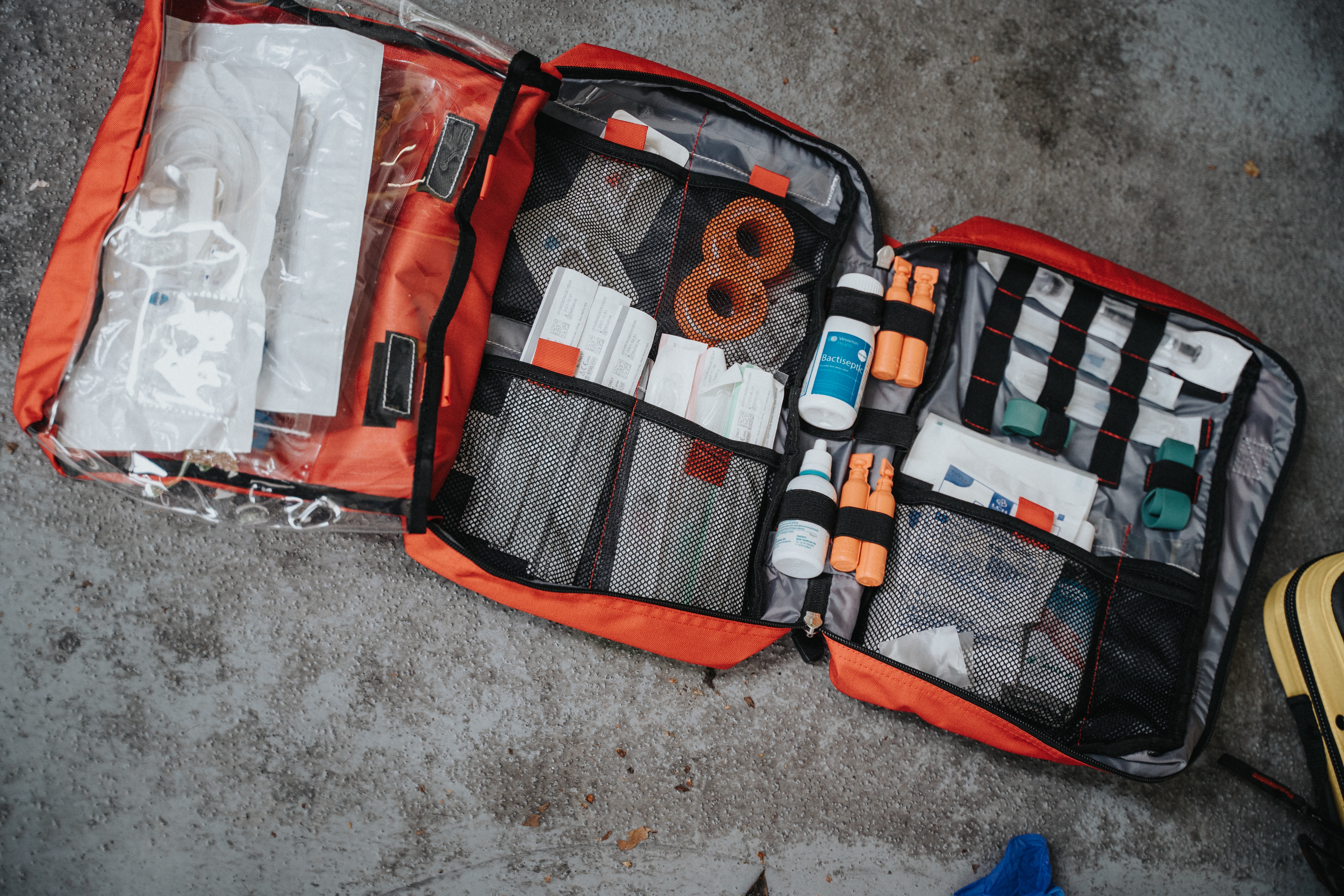
The main principle of treatment is to stop bleeding and restore the lost blood volume and includes the following steps:
- Bleeding control (requires rapid surgical haemostasis).
- Heating the patient.
- Infusion and transfusion therapy: the volume rate depends on the volume of blood loss - in severe hypotension, at least 100-150 ml/min (up to 500 ml/min in the presence of a self-saver).
- Speed control by blood pressure (BP), left ventricular diastolic filling pressure (LVDP) and central venous pressure (CVP).
If these parameters increase, the infusion rate should be reduced. For infusion, crystalloid solutions are used - 0.9% sodium chloride and 7.5-10% sodium chloride, as well as colloidal solutions, where hydroxy ethyl starch and modified gelatin, as well as the natural colloid albumin, are preferred.
Indications for haemotransfusions
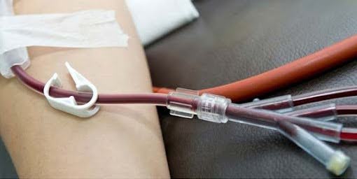
Hemotransfusion, or blood transfusion, is a procedure that involves the introduction of donor blood or its components into the recipient's blood vessels for therapeutic purposes. It is used to replace red blood cells and plasma proteins and to stop bleeding.
Hemotransfusion can be performed directly (from donor to recipient) or indirectly (donor blood is stored in a vial with a preservative before transfusion). It can be administered into the bloodstream by drip through peripheral (usually ulnar) or large veins, or in case of acute massive blood loss - intra-arterially.
Indications for blood transfusion:
- When the circulating blood volume is fully restored by fluids and the haemoglobin concentration is reduced to 70-80 g/l or lower, as well as in patients with concomitant diseases (coronary heart disease, heart failure, cerebrovascular disease or traumatic brain injury, respiratory failure) with a haemoglobin concentration of 90 to 100 g/l;
- If the hemoglobin concentration is ≥ 100 g/l, only uncontrolled profuse bleeding may be an indication for transfusion;
- In the case of refractory shock, cardiotonic and vasoconstrictor drugs should be used before volume therapy of shock (arterial hypotension with increased CVP and MAP);
- In case of refractory shock to the above therapy, glucocorticoid hormones may be used;
- In case of severe metabolic acidosis (pH < 7.1; BE < -10 mmol/l), isolation solutions (sodium bicarbonate) should be used;
- If you need pain relief, you should only use drugs that do not have a cardiovascular and vascular depressant effect;
- In case of inadequate breathing and the need for general anaesthesia, tracheal intubation and artificial lung ventilation should be performed;
- In the case of significant peripheral vasospasm, lactate acidosis, which do not disappear despite the restoration of circulating blood volume, increased blood pressure and CVP, it is necessary to use strictly dosed vascular dilators with a well-controlled effect (nitrates, α-adrenergic blockers) using infusomats;
- In the case of significant peripheral vasospasm, but in the presence of arterial hypotension, the use of cardiotonic agents or inodilators is indicated.


The result of treatment
Treatment outcome is a description of a person's condition after medical treatment and the duration of this treatment. The main goal of treatment is to completely stop bleeding, restore normal tissue perfusion, oxygen delivery and consumption, and the functioning of vital organs, such as consciousness, diuresis, lung gas exchange and the blood coagulation system.
After completion of treatment, the following results can be observed:
- Complete cessation of bleeding: An important indicator of the effectiveness of treatment is the complete cessation of bleeding, which ensures the restoration of normal blood circulation in the body.
- Restoration of normal tissue perfusion: This means that the blood delivers oxygen and other essential substances to the tissues efficiently enough to ensure their normal function.
- The functioning of vital organs: These organs include consciousness, diuresis (urination), gas exchange in the lungs and the blood clotting system.
Their normal functioning is a key indicator of successful treatment. The length of stay in the intensive care unit is usually 1-3 days, if there are no complications. However, this period may be extended in case of serious conditions or complications.
Possible complications and side effects
The post-shock period can lead to a number of complications and side effects that require close medical supervision and measures to manage them. Some of the most common complications and side effects include:
- Problems with the functioning of organs and systems: A variety of organ functioning problems can occur after shock, such as kidney, lung and liver problems. For example, kidney failure, impaired gas exchange in the lungs, or liver dysfunction may occur.
- Infections: After shock, a patient may become more susceptible to infections as their body may be vulnerable after a stressful period. Infections can affect various body systems and require adequate treatment.
- Bleeding from the stomach and intestines: This is another potential complication of shock. It can be caused by a variety of factors, including damage to the lining of the stomach or intestines due to stressful conditions or medication.
To prevent and effectively manage these complications, patients should receive intensive medical care for at least one day after recovery from shock. This is the only way to ensure that any potential problems that may arise during this critical period are properly identified and treated.
Recommendations for further treatment
After recovery from shock, it is important to provide patients with appropriate recommendations for further treatment and prevention of possible complications. Some recommendations include:
- Prevention of the development of multiple organ failure: Particular attention should be paid to preventing the development of multiple organ failure, as this can be a serious complication of shock. In particular, it is important to monitor the function of the lungs, kidneys, liver and blood coagulation system.
- Prevention of infectious complications: Post-shock patients may be more susceptible to infections due to a weakened immune system and the stress their bodies have been under. Therefore, it is important to take steps to prevent infections, including hygiene measures, vaccinations if necessary, and the use of antibiotics if needed.
- Prevention of bleeding from the stomach and intestines: Because bleeding from the stomach and intestines can be a serious complication of shock, it is important to take steps to prevent it. This may include limiting the use of drugs that can irritate the stomach, controlling blood pressure, and other measures to reduce the risk of bleeding.
The provision of these recommendations and their proper implementation can contribute to the patient's rapid recovery from shock and prevent the development of further complications.
Frequently asked questions and answers
What is haemorrhagic shock?
Haemorrhagic shock is an extremely serious condition that occurs when there is significant blood loss, when the body cannot provide enough blood circulation to
support vital organs.
What can be the causes of haemorrhagic shock?
Causes of haemorrhagic shock can include trauma, internal bleeding from ruptured blood vessels or organs, cutting, self-mutilation or fire, and certain medical conditions such as acute stomach or bowel ulcers.
What are the symptoms of haemorrhagic shock?
Symptoms of haemorrhagic shock may include pale skin, cold and damp sweat, rapid and shallow breathing, weakness, dizziness, and a fast pulse.