Combat chest injury, chest contusion
Content
- What is a chest contusion?
- Causes of slaughter
- Signs of slaughter
- Treatment of chest bruises
- What not to do with bruises
- What is combat thoracic trauma?
- Levels of care for thoracic trauma
- Recommendations for the treatment of thoracic injuries
- Surgical treatment of thoracic trauma
- Conclusion
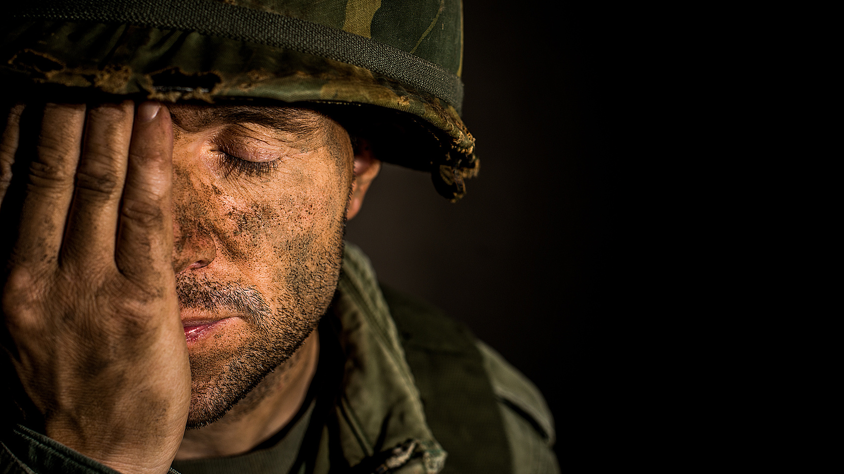
Combat-related chest injuries, including contusions, are a serious problem that requires immediate medical intervention. These injuries can have a significant impact on the functioning of the respiratory and cardiovascular systems, causing severe consequences for the health and life of the affected individuals. In this article, we will review the main medical treatment of such injuries, as well as provide recommendations for first aid and further treatment.
What is a chest contusion?
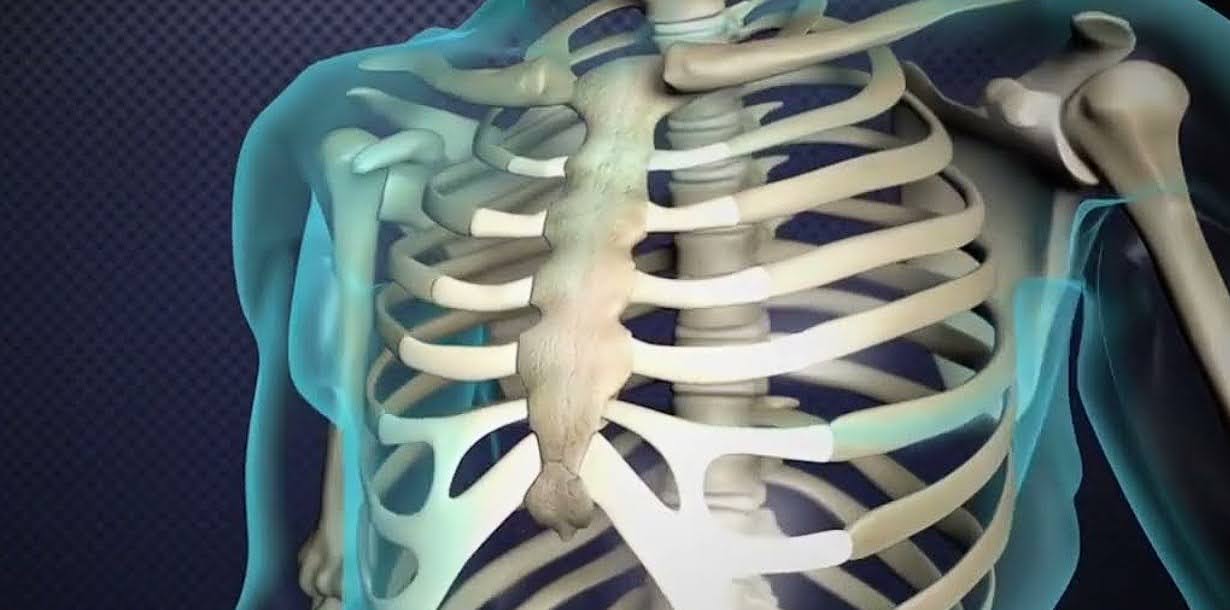
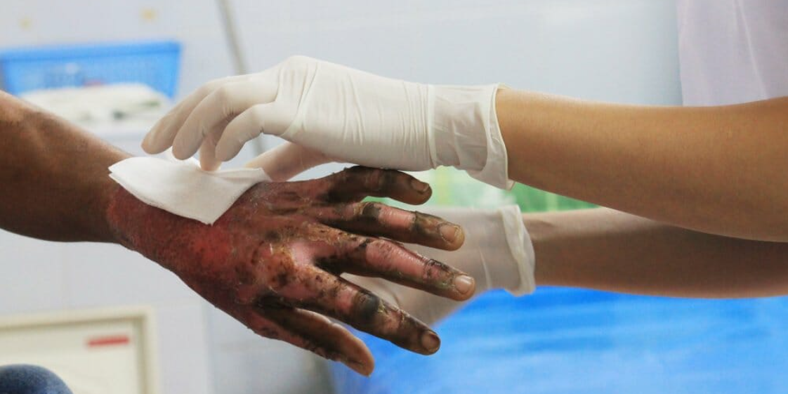
Chest contusion is a fairly common injury that occurs as a result of a significant blow to the chest or a fall on a sharp object. This injury involves a closed injury, in which internal organs can suffer serious damage.
The impact can cause damage not only to the surface tissues but also to important internal organs such as the lungs and heart. As a result of such a blow, the integrity of the skin and bone structures remains intact, but the chest is significantly compressed, which can lead to pain, swelling and bruising. Such contusions can often be complicated by various pathological conditions that require medical intervention to prevent serious consequences.
Chest injuries can vary in severity, from mild bruises to serious injuries that require immediate medical attention. In severe cases, a detailed examination, including X-rays or computed tomography, may be required to assess the condition of the internal organs and bones. Timely and proper treatment is key to preventing possible complications and ensuring a full recovery.
Causes of slaughter
Soft tissues are usually damaged when they hit a hard object or surface. For example, bruising of the back and tailbone when falling into water from a great height is quite common. Such injuries occur in a variety of situations, including combat injuries, sports and road traffic accidents, or domestic accidents.
The severity of slaughter depends on several key factors:
- The hardness of the injuring object. The harder the object, the more severe the injury. For example, a blow to a concrete surface will cause more serious injury than a soft object.
- The force and speed of the punch. Heavier and faster blows cause more damage. For example, a hard hit with a football can cause bruising that is different from that caused by a light blow.
- The size of the damage area. A large area of injury usually causes more complications than a small area. For example, a blow that involves a large area of soft tissue can lead to more swelling and haematoma.
- Tissue elasticity and blood supply. Tissues with good elasticity and blood supply can better withstand impacts and recover faster. For example, muscle tissue usually recovers faster than other tissues.
- The age of the victim. Younger people have more elastic tissue and better circulation, which contributes to a faster recovery. Older people may have less elasticity in their tissues and may take longer to recover.
Understanding these factors allows us to better assess the condition of victims and determine the best treatment methods to ensure their quick and complete recovery.
Signs of slaughter
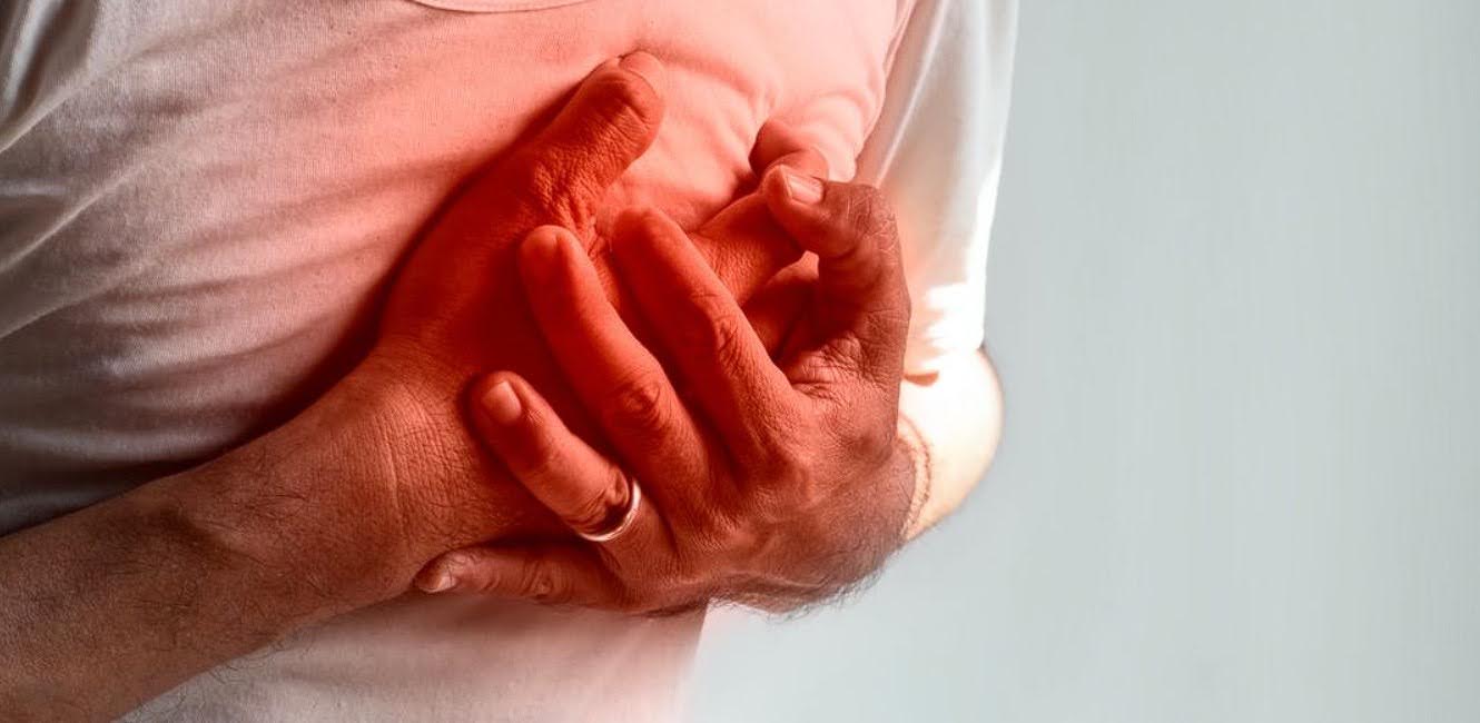
The main sign of a bruise is pain that occurs immediately at the moment of impact. A haematoma, an accumulation of blood in the tissues, may also appear. The larger the hematoma, the more painful the injury due to soft tissue compression and irritation of pain receptors.
External manifestations of contusion include swelling at the site of impact. In areas where there is more subcutaneous tissue, the swelling will be more noticeable. For example, bruises will be more pronounced on the face. When palpating the bruise site, the patient feels sharp pain. In case of limb injuries, their functions may be impaired.
Symptoms of rib contusions include increased pain when breathing, coughing or laughing. The absence of a characteristic crunch when the ribs are squeezed on both sides or in the anterior-posterior direction does not exclude a fracture.
Severe bruising of the chest can lead to other complications, such as damage to the heart or lungs. In such cases, heart and lung dysfunctions come to the fore.
Particular attention should be paid to joint contusions. In this case, haemorrhage can occur not only in the periarticular tissues, but also in the joint itself, which is called haemarthrosis. In this case, the joint is enlarged, its contours are smoothed out, and movements in it are difficult and painful. This form of contusion requires the prescription of special medicines.
Treatment of chest bruises
The general principles of treatment for all bruises remain the same. First of all, the affected area of the body needs rest. On the first day, cold compresses are recommended for 2-3 hours with half-hour breaks. This helps to reduce the size of the haematoma, reduce swelling and improve microcirculation at the site of injury.
On the second day, you can use ultrasound therapy. After the pain intensity decreases, compresses, ozokerite and paraffin applications are prescribed. It is also possible to use products that promote haematoma resorption, such as heparin ointments.
Electrophoresis with antibiotics or novocaine can be used for severe pain. Novocaine can also be used to perform a blockade if conventional painkillers are not effective enough.
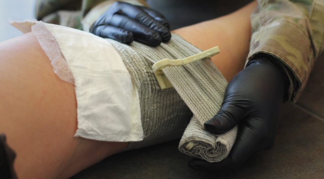
Large bruises after soft tissue contusions are usually punctured with a large needle, the blood is sucked out with a syringe, and then antibiotics are injected into the area and a sterile pressure dressing is applied.
If blood gets into the joint, it is also aspirated. After this procedure, a plaster cast is applied. Therapeutic exercise therapy (TET) is also prescribed for hemarthrosis.
What not to do with bruises
In the first day after slaughter, any thermal procedures on the affected area are strictly contraindicated. Warming causes the damaged vessels to dilate, which increases bleeding and leads to an enlargement of the haematoma.
High temperatures create a favourable environment for the growth of pathogens, which can lead to the development of phlegmon, a spillover suppuration of soft tissue. This inflammation is characterised by severe swelling, redness and pain, and may also be accompanied by fever and general weakness.
For the same reason, do not rub or massage a fresh bruise. Any mechanical irritation can worsen the condition of the injured area. In addition, if the injury is accompanied by a fracture that was not immediately detected, the sharp ends of the broken bone can further damage soft tissue, including blood vessels and nerve endings. This can lead to more serious complications, such as haemorrhage, nerve damage and the development of long-term pain.
This can not only increase the risk of phlegmon, but also contribute to the formation of larger haematomas. As a result, significant swelling can occur, making movement difficult and causing severe pain. In cases where a fracture is suspected, improper actions can lead to displacement of bone fragments, which will require more complex and lengthy treatment.
Thus, the first steps after a slaughter should be aimed at cooling the affected area and ensuring its rest. Cold compresses help to constrict blood vessels, reduce bleeding and swelling, and relieve pain. Adherence to these principles will help avoid complications and facilitate a faster recovery.
What is combat thoracic trauma?
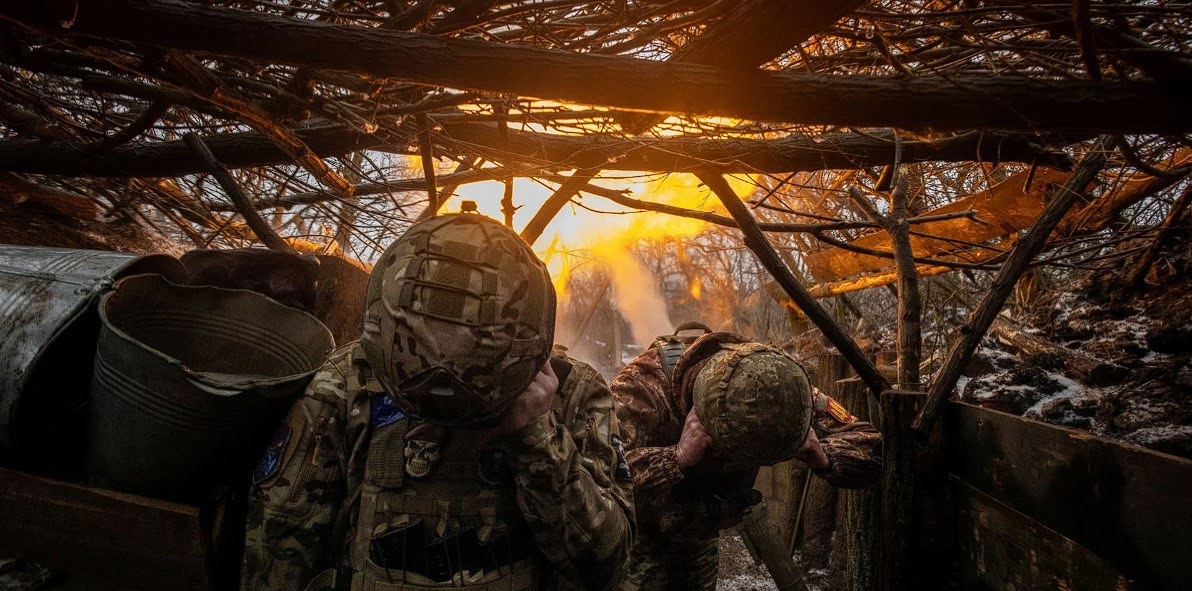
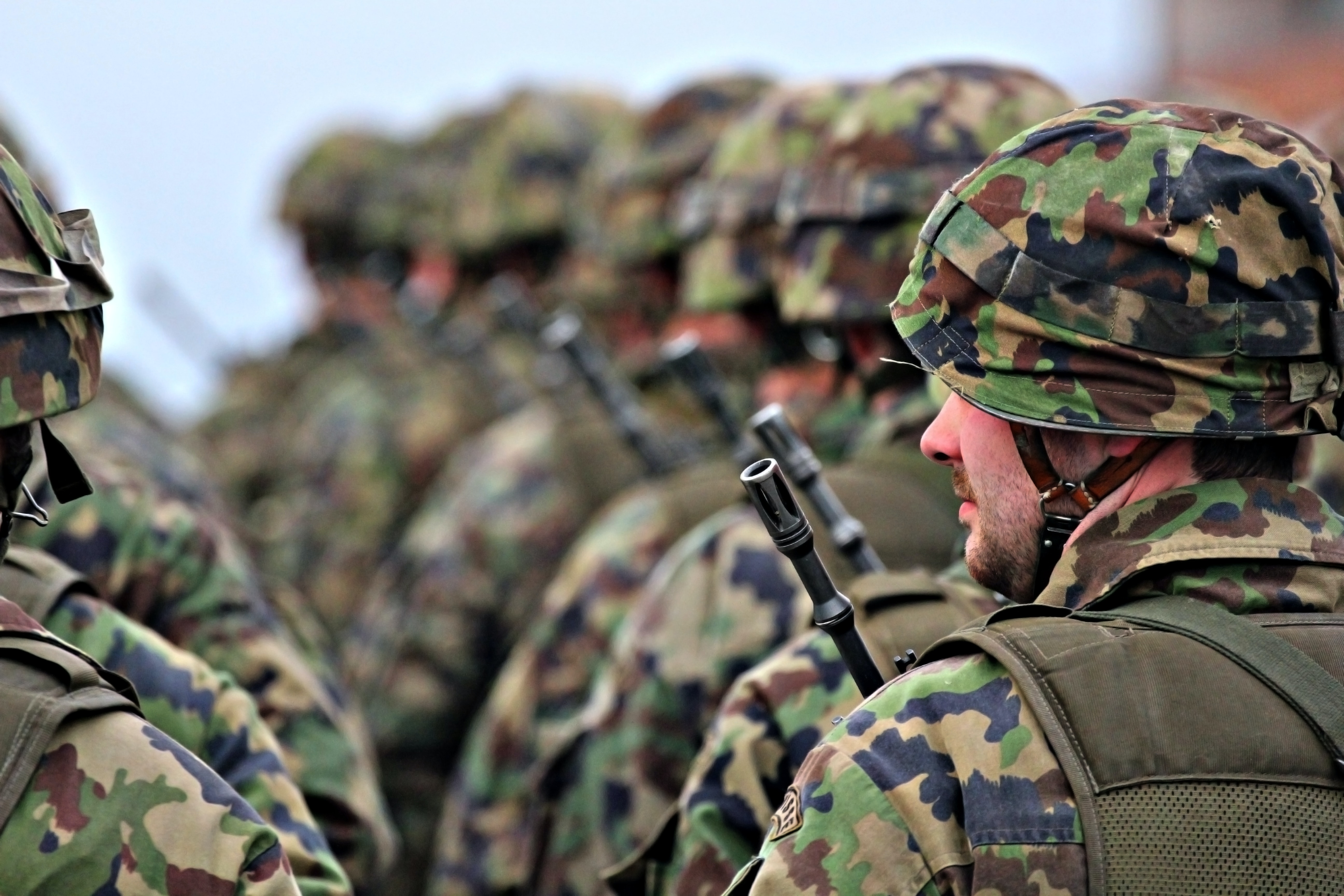
Combat thoracic trauma occurs as a result of injuries during military operations or conflicts. It can result from gunshot wounds, explosions, projectile fragments or other military mechanisms of trauma. Because combat environments often involve the use of firearms and other explosives, combat thoracic injuries can be severe and difficult to treat.
Thoracic injuries occur in 12-50% of civilian casualties. More than 90% of such injuries in civilians are caused by blunt force trauma, while surgical intervention is required in less than 10% of cases. In contrast, significant thoracic injuries are seen in only 10% of combat injured patients, but most of these are associated with penetrating trauma.
The most common thoracic injuries are pneumothorax, lung contusion and chest wall injury, with lung or large vessel injuries having the highest mortality rate. For the majority of patients with thoracic trauma, drainage of the pleural cavity (i.e., a "probe" thoracostomy) is recommended as the first diagnostic and therapeutic intervention.
In cases of more severe injuries, a thoracotomy (preferably anterolateral), sternotomy or other surgical procedures may be required depending on the condition of the injured.


Levels of care for thoracic trauma
When treating thoracic injuries, each level of care provides specific interventions adapted to their level of expertise and available resources. Thoracic injuries can be life-threatening, especially when they involve airway obstruction or tension pneumothorax. Such situations require immediate intervention, which increases the importance of efficient and reasonable resource allocation in healthcare facilities.
Here is a table detailing the measures at each level:
| Level | Description |
|---|---|
| Level I | The primary goal is immediate treatment or stabilization of thoracic injuries that pose a direct threat to life, such as airway obstruction and tension pneumothorax. When managing chest injuries, adherence to Tactical Combat Casualty Care guidelines is crucial. |
| Level II | Main focus is on resuscitation and surgical interventions to control injuries. Critical diagnostic and therapeutic measures include drainage of the pleural cavity. Patients with acute hemorrhage may require anterolateral thoracotomy or sternotomy. |
| Level III | For most injuries, definitive surgical procedures are recommended, except for complex tracheobronchial injuries or damage to major vessels. |
| Level IV | Radical therapy. |
This table outlines the different levels of care for thoracic trauma, from immediate response to life-threatening conditions to more complex procedures in more severe cases.
Recommendations for the treatment of thoracic injuries

Thoracic injuries are serious medical conditions that require competent and urgent treatment. These injuries, which involve the chest and surrounding structures, can occur for a variety of reasons, from motor vehicle accidents to falls or sports injuries.
Effective management and treatment of thoracic trauma is based on high medical expertise, prompt response and the use of modern diagnostic and treatment methods. Let's look at the basic principles and recommendations that will help medical staff to effectively manage thoracic trauma, ensuring the maximum possible rehabilitation and recovery of patients:
- For most patients with penetrating thoracic trauma and all patients with suspected significant pneumothorax or haemothorax, the first therapeutic intervention is recommended to be pleural drainage. In level I facilities, needle decompression is recommended for suspected tension pneumothorax.
- All patients with penetrating thoracic trauma or upper abdominal trauma are at risk of intrapericardial heart or large vessel injury, which can be detected by ultrasound or pericardial window.
- A left anterolateral thoracotomy (resuscitation thoracotomy) is the optimal approach for patients with penetrating thoracic trauma who are critically or extremely unstable, or with loss of vital signs on arrival. This procedure should be accompanied by drainage of the right pleural cavity or an extended thoracotomy incision (e.g., clamshell thoracotomy) if mediastinal or right chest injury is suspected.
- In patients with a pulsatile penetrating chest wound and haemopericardium, sternotomy is usually the preferred approach. If hemopericardium is uncertain, a subxiphoid pericardial window should be performed before sternotomy.
- The treatment of suspected subclavian vessel injuries is complex, technically demanding, and often involves a combination of sternotomy, anterior thoracotomy, and sub/ supraclavian incisions.
- For patients with suspected oesophageal perforation, tracheobronchial trauma or blunt trauma to large vessels, the primary focus should be on stabilisation, temporary control and extensive drainage rather than complete repair.
- In unstable patients with severe thoracoabdominal trauma, a combination of exploratory laparotomy with bilateral pleural drainage and a transdiaphragmatic pericardial window is recommended for the full assessment and treatment of severe injuries.
- Patients undergoing surgery for acute thoracic trauma should be placed in the supine position and the surgical field should be treated in the areas of potential incisions to control vital damage. A posterolateral thoracotomy in the setting of acute trauma is rarely justified.
- Even a minimal pneumothorax in patients who are transported for a long time is unacceptable. In cases of ambiguous clinical or radiological findings, the patient should be transported after draining the pleural cavity.
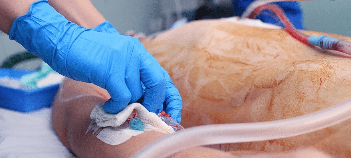
- In rare cases, such as complex thoracic trauma accompanied by severe hypoxia, hypercapnia or heart failure, extracorporeal membrane oxygenation (ECMO) may be required.
Surgical treatment of thoracic trauma
A patient with thoracic trauma who is scheduled for emergency surgery has a critically life-threatening injury. Therefore, it is recommended that the patient be placed in a supine position with the arms extended to the side, with the surgical field pretreated from the chin to the knees and from the chest to the elbows to ensure maximum accessibility for the necessary medical interventions.
A large-diameter access should be established (i.v., i.c., or central venous) and the airway secured with an endotracheal tube with cuff.
A left anterolateral thoracotomy, which is recommended in combination with right pleural drainage, is the optimal solution for all patients with penetrating thoracic trauma who are critically or extremely unstable.
In case of suspected intrapericardial trauma or bleeding in the right pleural space, the incision can be extended through the midline (i.e., a "grab" incision).
For patients with a pulsatile penetrating chest wound and haemopericardium, sternotomy is generally recommended as the optimal initial incision. If the diagnosis of haemopericardium is uncertain (e.g., if ultrasound is unavailable or equivocal, but cardiac injury is highly likely), a subxiphoid pericardial window should be performed before sternotomy.
The treatment of subclavian vascular injuries is complex. If the proximal portion of the left subclavian artery is suspected to be injured, a safe first incision that allows for proximal control is a left anterior thoracotomy in the third intercostal space.
The proximal control of the right subclavian artery is best achieved by means of a midline sternotomy. If necessary, intravascular balloon occlusion can be used to provide proximal control in more complex lesions.
After proximal control is achieved, the proximal area to the left or right of the middle part of the subclavian artery can be opened using a supraclavicular incision to repair the damaged subclavian artery.
The subclavian incision provides access to the more distal part of the subclavian artery and subclavian vein. In complex cases of subclavian vessel damage, a hatch incision provides maximum access.
When lung parenchyma is damaged, it is recommended that resection be performed in a non-anatomical manner, if possible. Anatomical resections should be performed only when the damage is not amenable to conservative treatment and involves the entire lobe that cannot be salvaged. Pneumonectomy should be performed only in cases of lung gate damage that does not respond to conservative treatment, as this procedure is associated with high mortality.
Posterolateral thoracotomy is rarely justified in the setting of acute trauma, as it limits access to neighbouring body cavities (such as the abdomen and neck) where injuries often occur. It should be considered only in cases of complete correction after ruling out or treating other injuries.
In patients with combined penetrating wounds to the chest and abdomen, as well as unstable haemodynamics, rapid insertion of bilateral chest tubes and a midline laparotomy with a pericardial window through the anterior diaphragm will allow for assessment of bleeding from all major body cavities.
If necessary, the laparotomy can be extended to a sternotomy to create a positive pericardial window. A separate anterolateral thoracotomy can also be performed in the event of bleeding into the pleural space. In the event of a diaphragmatic rupture, bleeding from the abdominal cavity may manifest as chest bleeding.
As described above, the principles and strategies of damage control are key in emergency thoracic surgery. Temporary haemostasis for chest wall defects or injuries can be achieved with gauze tamponade and haemostatic aids.
It is also possible to tamponade uncontrolled bleeding from the lungs, lung gates or thoracic aorta. However, pericardial tamponade is not recommended due to the risk of iatrogenic tamponade.
Following damage control procedures or if thoracic compartment syndrome is suspected during intensive care, temporary chest closure may be considered. This can be accomplished with negative pressure therapy or a simple adhesive dressing with a large-diameter chest drainage tube for drainage and aspiration.
Conclusion
Gunshot wounds and injuries pose a significant threat to people's lives, causing a variety of serious complications such as bleeding, airway damage, chest and lung injuries.
The effectiveness of treatment depends on the promptness of medical care and accurate diagnosis using modern methods such as computed tomography and ultrasound.
Early rehabilitation after an injury is important to reduce the risk of complications, including pneumonia. Preventive measures, such as vaccinations and the use of protective equipment, are recommended to reduce risks.